Overview
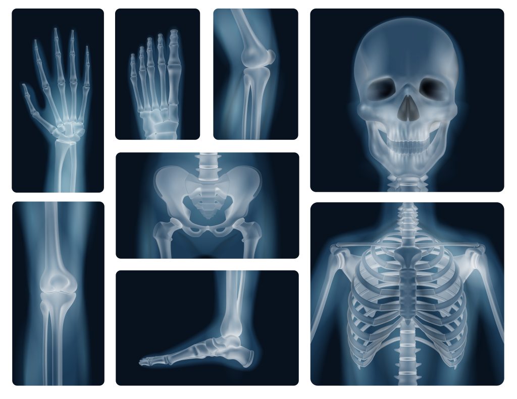
Digital radiography, another name for digital X-ray, is a contemporary medical imaging procedure that employs digital technology to acquire and display X-ray pictures. Digital X-rays, as opposed to conventional X-ray film, produce images that can be saved, examined, and edited on a computer.
PROCEDURE
The following steps are commonly included in the digital X-ray process:
- Preparation: As part of the preparation process, you will be asked to remove any clothing, jewellery, or other items that could obstruct the X-ray. A gown may be provided for you to wear.
- Positioning: The area of your body being inspected will be placed in front of the X-ray machine, and the X-ray beam will pass through it. To reduce image blurring, you could be requested to remain still and hold your breath for brief periods of time.
- Scanning: The region being investigated will be captured in one or more X-ray images by the X-ray machine. Typically, the entire process takes under ten minutes.
- Review: A radiologist will examine the digitally recorded images, analyse the findings, and deliver a report to your doctor.


Benefits of Digital X-Rays
Some of the key benefits of digital X-ray include the following:
- High-quality images: Images of substantially higher quality are produced by digital X-ray than by standard X-ray film. Radiologists can identify and treat a variety of ailments more easily thanks to the pictures’ improved clarity, sharpness, and detail.
- Efficiency: The ability to quickly save, view, and exchange digital X-ray images with other medical experts can speed up the diagnosis process and eliminate the need for extra X-ray scans.
- Environmentally friendly: digital X-ray images are produced and stored electronically, considerably reducing the environmental impact of the technology. This is in contrast to traditional X-ray film, which requires chemicals and other resources to develop.
- Advanced imaging techniques: Digital X-rays can produce detailed, three-dimensional images of the body that can be combined with other imaging techniques like CT scans and MRIs to help with the diagnosis and treatment of a variety of illnesses.
- Remote access: By enabling the long-distance transmission of pictures for radiologists to interpret, digital X-ray technology has been used to raise the standard of care in impoverished nations and remote locations.
- Dose modulation: Dose modulation: Dose modulation is a sophisticated imaging technique that can be used with digital X-ray technology to help patients receive less radiation exposure, particularly when they are children, pregnant women, or have frequent X-ray exams.
- Integration with other technologies: Digital X-ray systems can be integrated with other systems like PACS (picture archiving and communication systems), which aid in the archiving, retrieval, distribution, and presentation of medical pictures and data. This makes it easier to obtain and manage medical images, which improves the diagnostic procedure’s effectiveness.
- Reduced risk for patients: Patient danger is reduced because digital X-ray technology now allows X-rays to be taken of people who are unable to lie down or remain still, such as children or critically ill patients. This makes it possible for medical practitioners to take pictures of the body without endangering the patient.
Before beginning the test, the advantages and hazards should be carefully weighed because, like other medical imaging techniques, digital X-rays expose the patient to ionising radiation.
Machines Available For X-Rays At MSDC
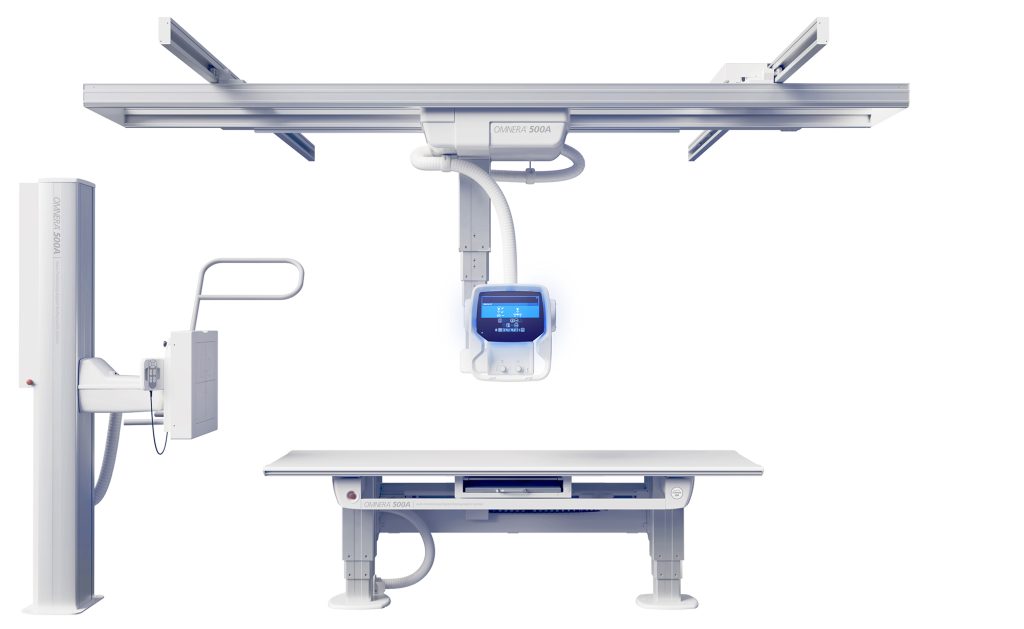
At MSDC, we employ the most recent imaging technologies to give our patients the finest care possible. The Siemens 500 mA X-ray machine, which is a high-end X-ray machine that generates high-quality images, is one of the technologies we use. The Fuji CR Device, a computerised radiography system that generates digital images, is another tool we employ. These two devices enable us to swiftly and effectively make photos of the highest quality.
The Siemens 500 mA X-ray machine is a potent imaging device that generates high-resolution images that are excellent for diagnosing conditions. It is a safe and efficient solution for our patients because it is made to deliver the highest-quality images with the least amount of radiation exposure. Advanced features on the machine, like automatic exposure control and automatic positioning, make it simple to use and help lower the possibility of human error.
The Fuji CR System, on the other hand, is a computerised radiography system that creates digital images. The image is captured using a specific plate by this device, which then sends it to a computer so that it can be viewed, saved, and shared. The Fuji CR System produces high-quality photographs, and it is also fitted with cutting-edge capabilities like automatic positioning and exposure management.
With the Fuji CR System and the Siemens 500 mA X-ray system, we can swiftly and effectively create high-quality images. These devices provide images that are easy to share with other medical experts and that can be used to diagnose a variety of illnesses. We have a skilled team of radiologists and technologists who are trained to use these tools, and they would be pleased to answer any questions you may have.


