Overview
A barium study is a type of medical imaging test that creates precise images of the digestive system using a contrast substance that contains the element barium. Barium sulphate, a white powder used as the contrast material, is combined with water to make a thick liquid that is either ingested or injected into the rectum. Barium highlights the digestive system’s organs and structures on X-rays, which makes it simpler for medical professionals to spot anomalies or issues.
Common Types of Barium Studies
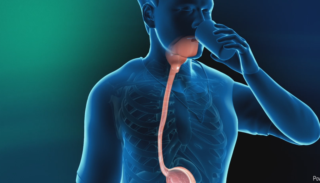
- Barium swallow: An upper gastrointestinal [UGI] study, often known as a “barium swallow,” is a test that looks at the oesophagus and upper stomach. A thick liquid called barium sulphate is consumed by the patient, who is then asked to stand in front of an X-ray machine while several pictures are taken as the barium passes through the digestive system. A barium swallow can be used to identify diseases including a hiatal hernia, ulcers, or oesophageal strictures.
PROCEDURE:
- To coat the lining of the oesophagus and stomach with barium sulphate, the patient is instructed to consume a thick liquid.
- As the barium travels through the digestive system, the patient is then positioned in front of an X-ray machine for a series of photographs to be obtained.
- The patient may be instructed to hold their breath and swallow at various points during the process in order to obtain particular images.
- Barium meal: This test is called a barium meal, often known as a small bowel study, and it looks at the small intestine. To assess the motility of the small intestine and its condition, the patient consumes the barium mixture and then undergoes an X-ray at predetermined intervals. It can aid in the diagnosis of diseases including inflammatory bowel disease and small intestinal polyps. It can aid in the diagnosis of diseases including inflammatory bowel disease and small intestinal polyps.
PROCEDURE:
- The patient is instructed to consume the barium meal.
- The patient will next be instructed to lie down and have X-rays at certain intervals.
- The patient may be asked to switch positions at some point during the treatment so that the entire small intestine may be seen.
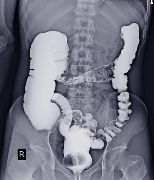
- Barium enema: The large intestine, commonly referred to as the colon, is examined with the barium enema, also known as a lower gastrointestinal [LGI] investigation. A tube is placed into the rectum while the patient is lying on a table. The colon is then filled with a mixture of barium and air, which is injected through the tube. As the barium fills the colon and draws the contour of the rectum, X-ray images are acquired. Inflammatory bowel illness, diverticulosis, and colon cancer can all be identified via a barium enema.
PROCEDURE:
- The patient is requested to lie down on a table for a barium enema, during which a tube is introduced into the rectum.
- The colon is then injected with a combination of air and barium using the tube.
- As the barium fills the colon and draws the contours of the colon and rectum, X-ray photographs are acquired.
- The patient could be requested to switch positions throughout the treatment so that the entire colon can be seen.
- Double-Contrast Barium Enema: This test is identical to the standard barium enema, but in addition to the barium, air is also injected into the colon during this procedure. This makes it possible to see the colon and rectum more clearly and precisely, which might be helpful in spotting polyps or other minor lesions that might be overlooked with a standard barium enema.
PROCEDURE:
- This technique is performed similarly to a standard barium enema, but air is also injected into the colon along with the barium.
- To provide a more precise and clear view of the colon and rectum, X-rays are taken after the colon has been filled with barium and air.
- CT-enteroclysis: To evaluate the small intestine, this procedure combines a CT scan with a barium swallow. Barium is used as a contrast agent, but CT scanning is used in place of conventional X-rays.
PROCEDURE:
- Similar to a barium swallow, a CT-enteroclysis involves the patient drinking a barium mixture before lying down for the scan.
- With this test, pictures of the small intestine are created using a CT scan and contrast material (barium).
- Following the procedure, the majority of the barium will be passed through the stools; however, some may pass through the urine. Water and other fluids should be consumed in large quantities to hasten this process. For a day or two after the test, it’s typical for the stools to be white or pale in colour.
The complete process typically takes between 30 and 60 minutes. The procedure could take longer, though, depending on the kind of test and the particular ailment being assessed. All barium studies are diagnostic tools; therefore, it’s crucial to keep in mind that which one is best for a given patient will depend on the patient’s unique symptoms and medical background. In each case, a doctor will consider the advantages and disadvantages of the test and will also go over the other diagnostic alternatives.


Why Barium Study test is done ?
The Barium Study test is used to assess and monitor gastrointestinal (GI) tract problems like:
- Oesophageal disorders: A barium swallow, also known as an upper gastrointestinal [UGI] examination, can be used to identify issues including a hiatal hernia, oesophageal strictures (narrowings), or reflux syndrome.
- Disorders of the gastrointestinal tract: A barium meal (or small bowel study) can be used to diagnose conditions like inflammatory bowel disease and small intestine tumours or polyps.
- Disorders of the colon: A barium enema (or lower gastrointestinal [LGI] study) can be used to identify issues including diverticulosis, inflammatory bowel disease, or colon cancer.
- Detecting polyps or other tiny lesions: A double-contrast barium enema can produce a more accurate and clear image of the colon and rectum, making it useful for finding polyps or other minor lesions that an ordinary barium enema could miss.
- Examining the small intestine: A CT-enteroclysis test is performed to assess the small intestine. It makes use of barium as a contrast agent but uses a CT scan rather than conventional X-rays.
Overall, barium tests are a crucial diagnostic tool for locating issues with the gastrointestinal tract and can be helpful in diagnosing disorders like hiatal hernias, ulcers, strictures, and malignancy. If you experience any symptoms that could point to a digestive system issue, speak with your doctor. You can then decide whether a barium study is the best course of action for you.


