Overview
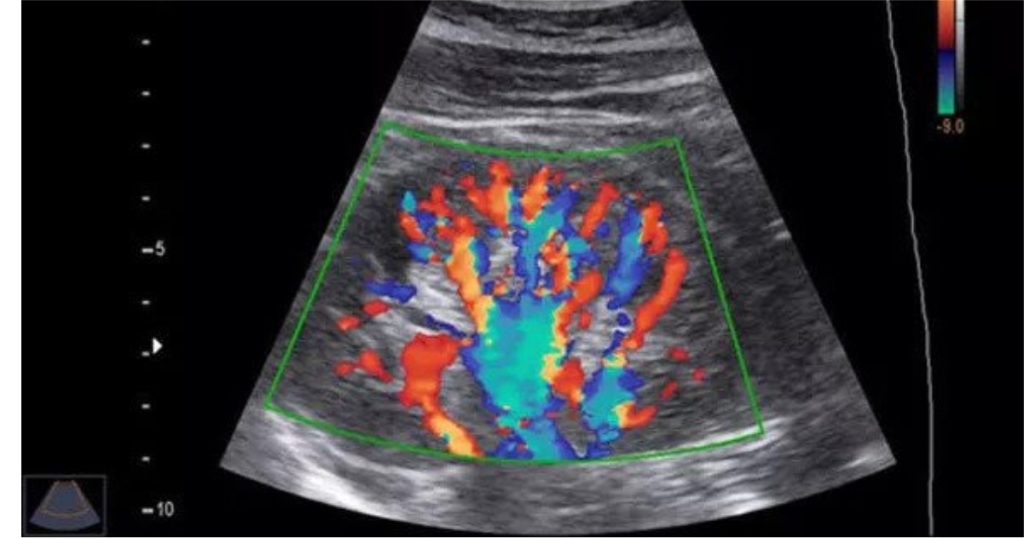
A non-invasive medical imaging method called Color Doppler ultrasonography employs high-frequency sound waves to provide finely detailed images of inside organs and blood vessels. The method is based on Doppler ultrasonography theory, which tracks variations in the frequency of sound waves as they return to the transducer.
WHY COLOR DOPPLER ULTRASOUND IS DONE ?
A variety of medical diseases, including the following, are diagnosed and monitored using a colour doppler:
- Blood Clots: Deep vein thrombosis, a disorder marked by blood clots in the legs’ deep veins, can be identified with colour Doppler ultrasound (DVT). Patients who have a high risk of DVT, such as those who have just undergone surgery or have a history of blood clots, can benefit most from this.
- Vascular Diseases: Various vascular illnesses, such as peripheral artery disease, carotid artery disease, and aortic aneurysms, can be detected and monitored using colour Doppler ultrasound. Additionally, it can be used to assess blood flow in the veins and find obstructions or irregular blood flow patterns.
- Pregnancy: Color Doppler ultrasound is frequently used throughout pregnancy to track the growth and development of the foetus. This technique can be used to visualise the foetus and placenta in great detail, as well as to identify any potential abnormalities, such as abnormal growth or placental difficulties.
- Abdominal Organs: Organs such as the liver, gallbladder, pancreas, and kidneys can be imaged using colour Doppler ultrasound. Additionally, it can be used to assess the blood flow in these organs, which can help with the identification of some illnesses like liver or kidney disease.
- Thyroid, Breast, and Testicles: Color Doppler ultrasonography can also be used to visualise the thyroid, breasts, and testicles. Diagnoses for illnesses like thyroid nodules, breast tumours, and testicular cancer may benefit from this.
- Guidance for biopsies: To make sure that samples are collected from the right place during biopsies, colour Doppler ultrasound can be used as a guide.

PROCEDURE
Color Doppler is a simple, painless, and safe treatment that normally takes 30-45 minutes to perform.
- Preparation: A color doppler ultrasound doesn’t require any extra preparation. During the treatment, you could be asked to remove any jewellery or clothing that might obstruct the imaging, as well as to wear a gown.
- Positioning: The area of your body being photographed will be covered with a transparent gel as you are positioned on an examination table. The gel is used to improve the clarity of the image and the transmission of sound waves through the skin.
- Transducer: The skin over the part of the body being scanned is covered with a tiny portable device known as a transducer. High-frequency sound waves from the transducer are sent into the body and returned to the transducer.
- Imaging: A computer analyses the reflected sound waves to provide fine-grained images of the interior organs and blood vessels. The radiologist will utilise these images, which are presented on a monitor, to assess the organs’ and vessels’ anatomy and functionality.
- Color Doppler: To assess the blood flow in the vessels, the colour Doppler method is used. It can be especially helpful in the detection of illnesses like deep vein thrombosis since it can find blockages or aberrant blood flow patterns (DVT).
- After the procedure: You’ll be able to leave and resume your regular activities after the procedure. The radiologist will review the photos before giving them to you or your referring doctor.
At our diagnostic center, advanced technology is used, and our technologists and radiologists are well qualified and experienced. Please get in touch with us if you have any additional questions or want to schedule an appointment for a colour Doppler ultrasound. Our staff will be more than delighted to serve you.


