Elastography
Overview
Elastography is a type of medical imaging that employs ultrasound to provide finely detailed pictures of the body’s interior organs. The method is founded on the idea that various tissue types have various elastic characteristics and that, by measuring these qualities, elastography can assist in the identification and diagnosis of a variety of disorders.
Types of ELASTOGRAPHY
There are two main types of elastography:
- Strain elastography: This technique produces images by capturing the movement of the tissue. An image is produced by using the tissue’s reaction to a modest amount of pressure applied by the ultrasonic probe. Stiffer regions of the tissue appear as bright spots on the picture, which illustrates the tissue’s relative elasticity. Strain elastography is mostly used to assess breast and liver tissue.
- Shear-wave elastography: This technique generates pictures using sound waves. An image is produced by using the tissue’s response to the sound waves that the ultrasonic probe sends into the tissue. Stiffer regions of the tissue appear as bright spots on the picture, which illustrates the tissue’s relative elasticity. Shear-wave elastography is used to assess a variety of body parts and systems, including the liver, breast, thyroid, prostate, and musculoskeletal system. It is more accurate than strain elastography.
Both methods of elastography are non-invasive, use no ionising radiation, and are generally safe and well-tolerated medical procedures. Repeated scans are also possible without worrying about radiation exposure over time.
Uses of ELASTOGRAPHY
A variety of body organs and systems, including the liver, breast, thyroid, prostate, and musculoskeletal system, can be examined with elastography. It can be used to find a number of things, like:
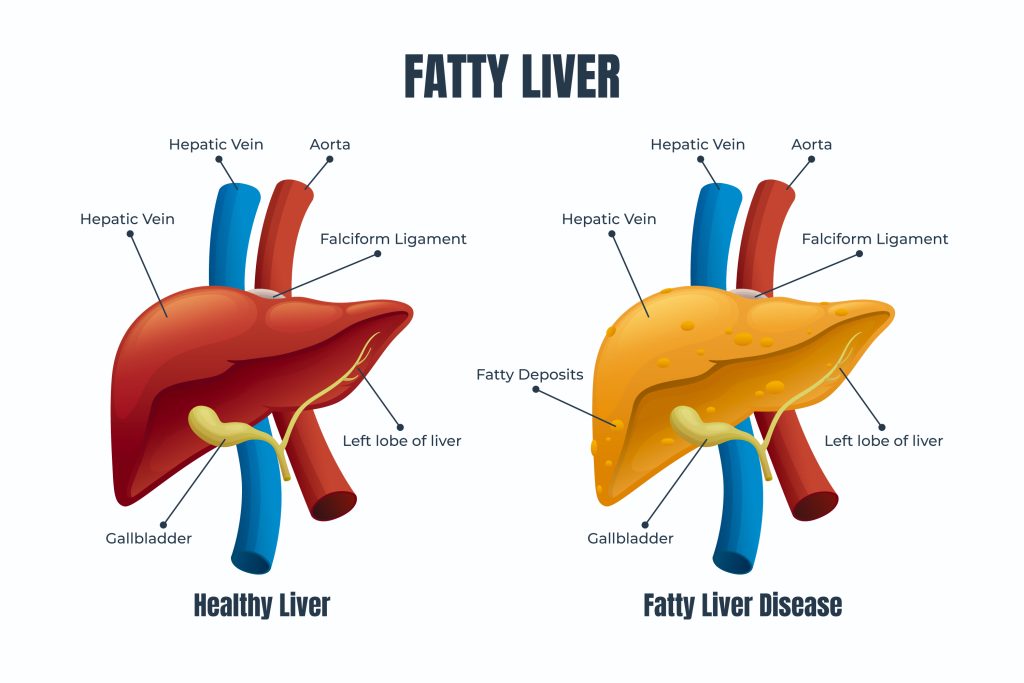
- Liver diseases: Liver fibrosis and cirrhosis can be detected and graded using elastography, which can assist in identifying people at risk of liver failure.
- Breast cancer: By using elastography to distinguish between benign and malignant breast lesions, fewer unnecessary biopsies may be performed.
- Disorders of the thyroid: Elastography can be used to distinguish between benign and malignant thyroid nodules, thereby minimising the need for pointless biopsies.
- Prostate cancer: Elastography can be used to distinguish between benign and cancerous prostate lesions, which can help cut down on the number of pointless biopsies.
- Musculoskeletal Disorders: Tendinopathies, as well as muscle and ligament injuries, can all be detected and graded according to their severity using elastography.

PROCEDURE
The following steps are commonly included in the elastography procedure:
- Preparation: You’ll be asked to take off any clothing or jewellery that could get in the way of the ultrasound during preparation. A gown may be provided for you to wear.
- Positioning: The part of your body being inspected will be cleansed and coated with a gel to let the ultrasound waves travel through your skin when you are positioned in front of the ultrasound equipment.
- Scanning: The ultrasonic probe will be positioned on your skin and moved over the area being evaluated during the scanning process. The probe will produce sound waves, and the response of the tissue to these waves will be measured.
- Image creation: Using the information gathered by the probe, an image of the tissue will be produced and shown on a computer screen. Stiffer regions of the tissue appear as bright spots on the picture, which illustrates the tissue’s relative elasticity.
- Interpretation: A radiologist or other healthcare professional will study and interpret the image and use the results to identify and diagnose any medical concerns.
Depending on the size of the area being investigated and the type of elastography being used, the full process typically takes 30 to 60 minutes. If you are pregnant or think you might be pregnant, or if you have any allergies or medical issues, it’s crucial to let the radiologist know. Although elastography is thought to be painless and safe, occasionally it can be uncomfortable.
BENEFITS
Some of the key advantages of elastography include the following:
- Non-invasive: Since elastography does not involve the use of ionising radiation, it is a non-invasive procedure that is generally safe and well-tolerated by patients. Repeated scans are also possible without worrying about radiation exposure over time.
- Accurate diagnosis: A variety of illnesses, including liver fibrosis and cirrhosis, breast cancer, thyroid problems, prostate cancer, and musculoskeletal ailments, can be found and diagnosed using elastography.
- Reduced need for biopsies: Biopsies are less frequently required because elastography can distinguish between benign and malignant tumors, which can help reduce the number of unnecessary biopsies.
- Improved patient care: By recognising and diagnosing medical issues earlier, elastography, which may be used to analyse a variety of body parts and structures, can assist in enhancing patient care.
- Cost-effective: Elastography is a cost-effective imaging technology since it can eliminate the need for other expensive imaging modalities and does not require any additional materials or chemicals for image development.
- Remote access: By enabling the long-distance transmission of pictures for radiologists to analyse, elastography technology has been used to raise the standard of care in remote and underdeveloped nations.
- Advanced imaging techniques: By combining elastography with other imaging techniques like CT scans and MRI, detailed, three-dimensional images of the body may be created. These images can be extremely helpful in the diagnosis and treatment of a variety of illnesses.
It’s crucial to understand that elastography cannot take the place of other imaging modalities and that the results of this test should be evaluated in conjunction with those of other clinical exams and imaging tests.


