Overview
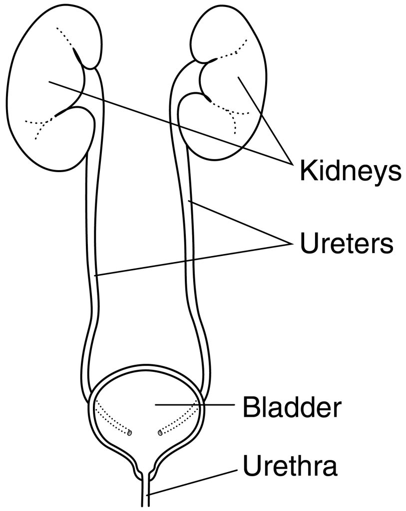
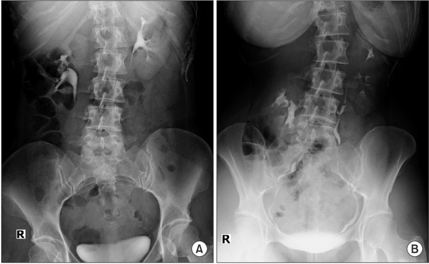
Intravenous Urogram (IVU) is a diagnostic imaging procedure that creates detailed images of the kidneys, ureters, and bladder using an X-ray and contrast dye. It is a non-invasive method that can assess kidney function, detect and diagnose a number of disorders affecting the urinary system, and track the success of treatment for specific urinary tract conditions.
CONDITIONS DIAGNOSED VIA IVU
IVU is used to identify a number of urinary tract disorders. The following are a few of the conditions that IUP can identify:
- Kidney stones: IVU is able to identify the presence of kidney stones as well as their size and location. The optimal course of treatment can be chosen using this information.
- Blockages in the urinary tract: IVU is able to identify obstructions in the urinary tract, such as bladder blockages or narrowing of the ureter. This information is crucial in figuring out what causes symptoms like frequent urinary tract infections or difficulties peeing.
- Tumors or cysts in the kidneys or bladder: IVU has the ability to identify the existence of tumours or cysts in the kidneys or bladder. The type, stage, and ideal course of treatment can all be determined with the help of this information.
- Ureteropelvic junction obstruction: It is a blockage at the junction of the ureter and the renal pelvis that can be found via IVU. A kidney stone, a congenital anomaly, or an enlarged prostate are possible causes.
- Reflux of urine into the kidneys: Urine moving backward from the bladder into the ureters and kidneys is referred to as vesicoureteral reflux, and it can be identified via an IVU test. This can eventually harm the kidneys and result in recurring urinary tract infections.
- Hydronephrosis: IVU can identify the dilation of the renal pelvis and calyces, known as hydronephrosis. This may be brought on by a congenital anomaly or a blockage in the urinary tract, such as a kidney stone or tumor.
- Hydroureter: IVU can identify the condition known as hydroureter, which is characterized by ureteral dilation. This may be brought on by a congenital disorder or a blockage in the urinary tract, such as a kidney stone or tumor.


PROCEDURE
The procedure normally takes 30 to 60 minutes to complete and is done in a hospital or diagnostic imaging facility. The specific steps of the IVU process are listed below:
- Preparation: The patient will be given instructions on how to prepare for the treatment, including how long they should fast and how much water they should drink to help flush out their urinary system. Additionally, the patient will be asked to take off any jewellery or other metallic items that could obstruct the X-ray images.
- Contrast dye injection: The process starts with the injection of a contrast dye into an arm vein. Typically, a syringe and a tiny needle are used to inject the dye.
- Waiting for the dye to reach the kidneys: After the injection, the patient will be advised to wait for a specific amount of time, typically between 10 and 15 minutes, for the dye to reach the kidneys. The patient can be asked to urinate during this period in order to let the urinary system flush out.
- X-ray images: Once the dye has reached the kidneys, X-ray images will be taken at timed intervals as the dye moves through the urinary tract. The patient would be told to stay still while he or she was being x-rayed while lying on an exam table.
- Post-operation: The patient might be asked to wait for a short while after the procedure to make sure there have been no negative reactions to the contrast dye. The patient could feel a little uncomfortable or taste something metallic in their mouth, but these symptoms typically go away in a few hours.
- Review of the images: The radiologist will look through the images to find any obstructions or other issues, as well as any abnormalities in the kidneys, ureters, or bladder’s size, shape, or location. The IVU results will be provided to the referring physician, who will then diagnose the patient using them along with other diagnostic procedures and clinical data.

ADVANTAGES
The following are a few benefits of IVU:
- High level of accuracy: IVU is a very accurate diagnostic tool that may identify and classify a variety of urinary tract problems, including kidney stones, obstructions in the urinary tract, tumours or cysts in the kidneys or bladder, and obstruction of the ureteropelvic junction.
- Non-invasive: IVU does not involve making any incisions or undergoing any type of surgery. Patients normally tolerate it well, and there is little chance of problems.
- Provides detailed images: IVU produces comprehensive images of the kidneys, ureters, and bladder using X-ray and contrast dye. This lets the radiologist see the size and shape of the organs, as well as any problems or abnormalities.
- Evaluation of kidney function: IVU can measure how quickly and how much the contrast dye leaves the body through the urine to determine how well the kidneys are functioning.
- Monitoring the effectiveness of treatment: IVU is a tool that can be used to keep track of how well treatment is working for conditions that affect the urinary tract, like kidney stones and blockage of the ureteropelvic junction.
- Cost-effective: IVU is a diagnostic tool that can be used without the need for costly equipment or specific training.
Other Service
Need help ?
Expertise and advanced technology are both essential for better diagnosis of the body. Contact us if you require any assistance with your diagnostics.


